


Sarcomere Structures: The Ultimate Guide to Muscle Contraction
The human body is a marvel of engineering, and at the heart of its movement lies a fascinating cellular structure: the sarcomere. These tiny, repeating units are responsible for the contraction of muscle fibers, enabling everything from walking and lifting to breathing and blinking. Understanding sarcomere structures is crucial for anyone interested in anatomy, physiology, exercise science, or even the treatment of muscular disorders. This comprehensive guide will delve into the intricacies of the sarcomere, exploring its components, function, and significance in the human body.
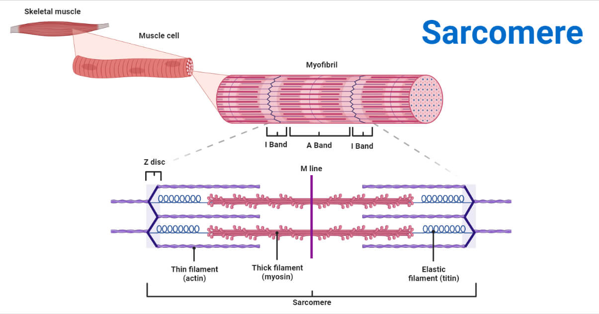
What is a Sarcomere? The Basic Building Block
A sarcomere is the fundamental contractile unit of a muscle fiber. It’s a highly organized arrangement of protein filaments that slide past each other, causing muscle contraction. Think of it as the smallest functional unit responsible for generating force and movement. These units are arranged end-to-end within a muscle fiber, forming long chains that contribute to the overall muscle’s ability to contract.
Key Components of the Sarcomere: A Detailed Breakdown
The sarcomere is composed of several key protein filaments and structures that work in concert to facilitate muscle contraction. Let’s break down the essential components:
- Z-Discs (Z-Lines): These are the boundaries of the sarcomere, acting as anchors for the thin filaments. They are made primarily of the protein alpha-actinin.
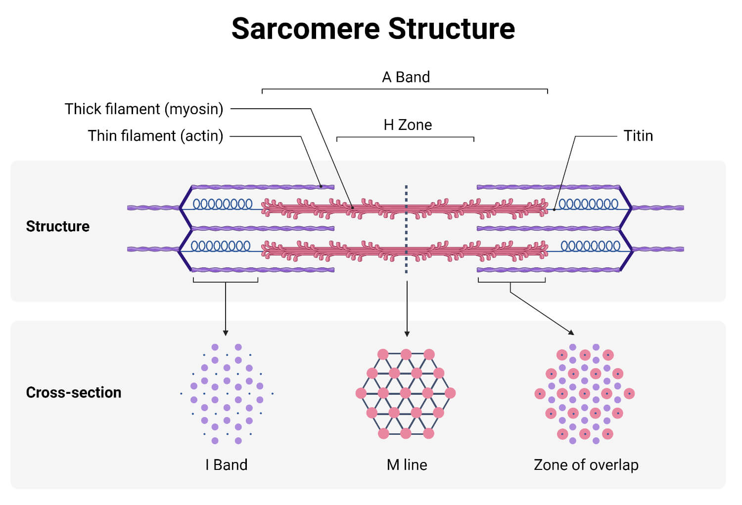
- I-Band (Light Band): This region contains only thin filaments (actin) and extends from the Z-disc to the beginning of the thick filaments. It appears lighter under a microscope.
- A-Band (Dark Band): This region contains both thick filaments (myosin) and the overlapping portions of thin filaments. It appears darker under a microscope.
- H-Zone: Located in the middle of the A-band, this region contains only thick filaments (myosin).
- M-Line: This line runs down the center of the H-zone and helps to hold the thick filaments in place. It’s composed of proteins that connect the myosin filaments.
- Thin Filaments (Actin): These filaments are primarily composed of the protein actin, along with regulatory proteins like tropomyosin and troponin. Actin filaments have binding sites for myosin.
- Thick Filaments (Myosin): These filaments are made up of the protein myosin, which has a globular head that binds to actin and a tail that forms the body of the filament. The myosin heads contain ATPase, an enzyme that hydrolyzes ATP to provide energy for muscle contraction.
The Mechanism of Muscle Contraction: Sliding Filament Theory
The process of muscle contraction is explained by the sliding filament theory. This theory proposes that muscle contraction occurs when the thin and thick filaments slide past each other, causing the sarcomere to shorten. Here’s a step-by-step overview:
- Nerve Impulse: A nerve impulse (action potential) reaches the neuromuscular junction, the site where a motor neuron connects to a muscle fiber.
- Calcium Release: The nerve impulse triggers the release of calcium ions (Ca2+) from the sarcoplasmic reticulum, a specialized network of tubules within the muscle fiber.
- Troponin Activation: Calcium ions bind to troponin, a protein associated with the thin filament. This binding causes a conformational change in troponin, which, in turn, moves tropomyosin, another protein that covers the myosin-binding sites on actin.
- Myosin Binding: With the myosin-binding sites on actin exposed, myosin heads can bind to actin, forming cross-bridges.
- Power Stroke: The myosin heads pivot, pulling the thin filaments towards the center of the sarcomere (the M-line). This is the “power stroke” that shortens the sarcomere. This process is driven by the hydrolysis of ATP by the myosin head.
- Detachment and Reattachment: ATP binds to the myosin head, causing it to detach from actin. The myosin head then hydrolyzes the ATP, returning to its “cocked” position and ready to bind to another actin site further along the thin filament.
- Sarcomere Shortening: This cycle of binding, power stroke, detachment, and reattachment repeats, causing the sarcomere to shorten, and the muscle fiber to contract.
- Muscle Relaxation: When the nerve impulse stops, calcium ions are pumped back into the sarcoplasmic reticulum. The troponin-tropomyosin complex returns to its original position, blocking the myosin-binding sites on actin, and the muscle relaxes.
The Role of Sarcomeres in Different Muscle Types
Sarcomeres are the fundamental building blocks of striated muscle, which includes both skeletal muscle (responsible for voluntary movements) and cardiac muscle (responsible for pumping blood). Smooth muscle, found in the walls of internal organs, does not have a sarcomere structure; its contraction mechanism is different. The arrangement and characteristics of sarcomeres vary slightly depending on the muscle type and its specific function, but the core principles of contraction remain the same.
Implications of Sarcomere Structure for Exercise and Health
Understanding sarcomere structure is crucial for understanding how muscles respond to exercise and how to optimize muscle performance.
- Muscle Hypertrophy: Resistance training can lead to muscle hypertrophy, an increase in muscle size. This occurs, in part, due to the addition of new sarcomeres in parallel, increasing the muscle’s ability to generate force.
- Muscle Atrophy: Conversely, lack of use (e.g., during prolonged bed rest) can lead to muscle atrophy, a decrease in muscle size. This is often associated with a loss of sarcomeres.
- Muscle Injury and Disease: Sarcomere dysfunction can be a factor in various muscular disorders, such as muscular dystrophy. Research into sarcomere structure is ongoing to find solutions for these diseases.
Conclusion: The Power of the Sarcomere
The sarcomere is a remarkable structure, the engine behind our ability to move, breathe, and live. By understanding its components and the process of muscle contraction, we gain a deeper appreciation for the complexity and efficiency of the human body. Whether you’re a student, athlete, or simply curious about how your body works, the knowledge of sarcomere structures is invaluable.
Frequently Asked Questions (FAQs)
1. What is the difference between the I-band and the A-band?
The I-band contains only thin filaments (actin), while the A-band contains both thick filaments (myosin) and the overlapping portions of thin filaments. The I-band appears lighter under a microscope, while the A-band appears darker.
2. What role does calcium play in muscle contraction?
Calcium ions (Ca2+) are essential for muscle contraction. They bind to troponin, causing a conformational change that moves tropomyosin, exposing the myosin-binding sites on actin.
3. What is the role of ATP in muscle contraction?
ATP (adenosine triphosphate) provides the energy for muscle contraction. It is used by the myosin heads to detach from actin and to return to their “cocked” position, ready for another cycle of binding and power stroke.
4. How does the sliding filament theory explain muscle contraction?
The sliding filament theory states that muscle contraction occurs when the thin (actin) and thick (myosin) filaments slide past each other, causing the sarcomere to shorten. This is achieved through the repeated binding, power stroke, detachment, and reattachment of myosin heads to actin filaments.
5. Are sarcomeres present in all types of muscle?
No, sarcomeres are found in striated muscles (skeletal and cardiac). Smooth muscle, which lines the walls of internal organs, does not have a sarcomere structure.
