


The Sarcomere: A Beautiful, Labeled Puzzle of Muscle Contraction
The human body is a marvel of engineering, and at the heart of its incredible movements lies the humble sarcomere. This microscopic structure, the fundamental unit of muscle contraction, is a complex and exquisitely organized system. It’s a beautiful, labeled puzzle where each piece plays a crucial role in enabling us to walk, run, lift, and even breathe. Understanding the sarcomere is key to unlocking the secrets of muscle function and how our bodies achieve such remarkable feats of movement.
This article delves into the intricate workings of the sarcomere, exploring its components, the process of contraction, and its significance in health and disease. Prepare to be amazed by this tiny but mighty component of life!
The Anatomy of a Tiny Powerhouse: Sarcomere Structure
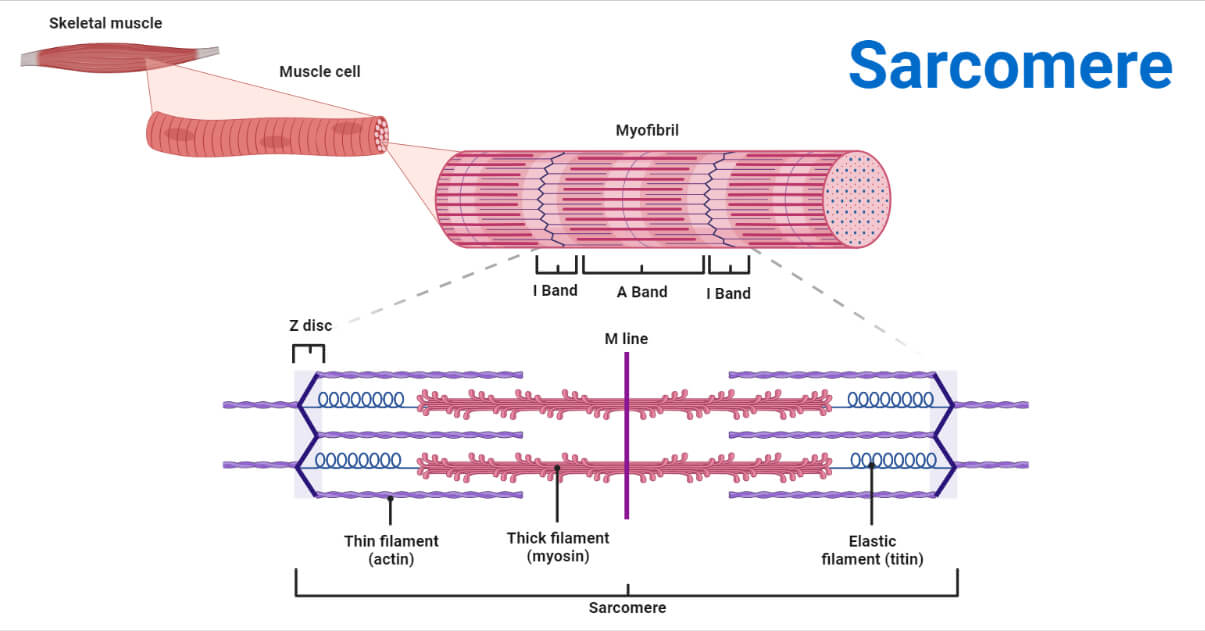
The sarcomere is a highly organized arrangement of protein filaments. It’s essentially a repeating unit within a muscle fiber, responsible for the muscle’s ability to shorten and generate force. Let’s break down the key players:
- Z-discs (Z-lines): These are the boundaries of the sarcomere. They are dense protein structures that anchor the thin filaments (actin).
- M-line: Located at the center of the sarcomere, the M-line anchors the thick filaments (myosin).
- A-band: This band encompasses the entire length of the thick filaments and includes the overlapping region of thick and thin filaments.
- I-band: This band contains only thin filaments and is located on either side of the Z-discs.
- H-zone: This is the central region of the A-band where only thick filaments are present, with no overlap with thin filaments at a relaxed state.
- Thick Filaments (Myosin): These are composed of the protein myosin, which has heads that bind to actin and generate force.
- Thin Filaments (Actin): These are composed of the protein actin, along with regulatory proteins like tropomyosin and troponin.
- Tropomyosin: A protein that wraps around actin filaments, covering the binding sites for myosin.
- Troponin: A protein complex that binds to tropomyosin, actin, and calcium ions.
The Sliding Filament Theory: How Contraction Happens
The magic of muscle contraction is explained by the sliding filament theory. This theory describes how the thick and thin filaments slide past each other, shortening the sarcomere and ultimately causing the muscle to contract. The process unfolds in a series of coordinated steps:
- Nerve Impulse: A signal from a motor neuron arrives at the neuromuscular junction, triggering the release of acetylcholine.
- Calcium Release: Acetylcholine initiates a cascade of events that leads to the release of calcium ions (Ca2+) from the sarcoplasmic reticulum (SR), an intracellular storage site.
- Troponin’s Role: Calcium ions bind to troponin, causing it to change shape. This shift in troponin moves tropomyosin away from the myosin-binding sites on the actin filaments, exposing them.
- Cross-bridge Formation: Myosin heads bind to the exposed actin binding sites, forming cross-bridges.
- Power Stroke: The myosin heads pivot, pulling the actin filaments towards the center of the sarcomere (the M-line). This “power stroke” shortens the sarcomere.
- Detachment and Reattachment: ATP (adenosine triphosphate) binds to the myosin heads, causing them to detach from actin. The ATP is then hydrolyzed (broken down) into ADP (adenosine diphosphate) and phosphate, re-energizing the myosin heads and preparing them for another cycle.
- Muscle Relaxation: When the nerve impulse stops, calcium ions are pumped back into the SR. Troponin and tropomyosin return to their original positions, blocking the myosin-binding sites on actin. The muscle relaxes.
The Sarcomere in Health and Disease
The sarcomere’s proper function is critical for overall health and mobility. Disruptions in the sarcomere’s structure or function can lead to various muscle disorders:
- Muscular Dystrophies: These genetic disorders involve progressive muscle weakness and degeneration due to defects in proteins involved in sarcomere structure (e.g., dystrophin in Duchenne Muscular Dystrophy).
- Myopathies: These are disorders that directly affect the muscle fibers. They can result from a variety of factors, including genetic mutations, infections, or inflammatory conditions.
- Sarcopenia: Age-related loss of muscle mass and strength, which often involves changes in sarcomere structure and function.
Understanding the sarcomere is crucial for developing treatments for these debilitating conditions. Research continues to focus on understanding the molecular mechanisms of sarcomere dysfunction and identifying potential therapeutic targets.
The Sarcomere: A Continuing Discovery
The sarcomere, though microscopic, holds enormous significance for our physical well-being. It is a testament to the intricate design of the human body and its ability to generate movement. As we continue to unravel the complexities of the sarcomere, we gain a deeper appreciation for the wonders of biology and the potential to improve human health.
Frequently Asked Questions (FAQs)
1. What is the primary function of the sarcomere?
The primary function of the sarcomere is to contract, enabling muscle fibers to shorten and generate force, leading to movement.
2. What is the role of calcium in muscle contraction?
Calcium ions bind to troponin, which causes tropomyosin to move, exposing the myosin-binding sites on actin. This allows myosin heads to bind to actin and initiate the power stroke, which results in muscle contraction.
3. What is the difference between the A-band and the I-band?
The A-band encompasses the entire length of the thick filaments (myosin) and includes the overlapping region with the thin filaments (actin). The I-band contains only thin filaments and is located on either side of the Z-discs.
4. How does ATP contribute to the muscle contraction process?
ATP is required for the detachment of the myosin heads from the actin filaments and the re-energization of the myosin heads, allowing them to bind to actin and repeat the cycle of contraction.
